Figure 3.
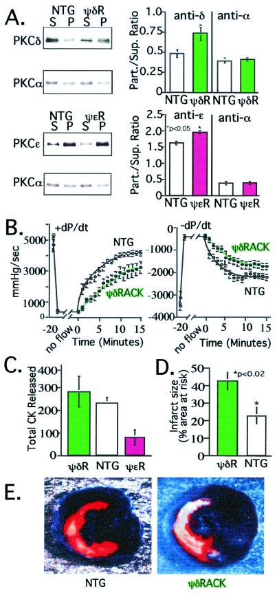
ψδRACK transgenic mice exhibit increased damage by cardiac ischemia. (A) Western blot and analysis of PKC distribution in cytosolic and particulate fractions from ψδRACK mice (ψδR, Upper; green bars) and ψɛRACK transgenic mice (ψɛR, Lower; magenta bars) and their nontransgenic littermates (NTG; white bars) was carried out as described (13) to demonstrate isozyme-selective translocation of PKC. (Right) Histogram shows mean ± SEM of data from eight mice for each group. (B) Hemodynamic parameters were monitored in hearts from transgenic ψδRACK transgenic mice (green symbols) and their nontransgenic littermates (white symbols) after global ischemia. Left ventricular pressure and real time derivative was monitored via a catheter placed in the ventricular apex (13). Hemodynamic measurements were recorded every 20 sec throughout reperfusion. Data are mean ± SEM of 12 ψδRACK and 11 nontransgenic mice. (C) Fractions of perfusate from mice used in B were collected throughout reperfusion and CK activity was assessed to determine cell damage. For comparison, data from ψɛRACK mice are also included. Data are mean ± SEM of six ψδRACK mice (green bar), six nontransgenic mice (white bar), and seven ψɛRACK mice (magenta bar). (D) Infarct size as a percent of the region of risk in mice with sustained δPKC activation (ψδRACK mice; green bar) and nontransgenic littermates (white bar) was determined in vivo after coronary occlusion followed by 24 h of reperfusion as described (mean ± SEM) (22, 23). The area at risk was not significantly different between the nontransgenic and ψδRACK mice (36 ± 3% and 41 ± 5% of left ventricle for nontransgenic and ψδRACK mice, respectively). Data are from eight 30- to 34-week-old transgenic females and five nontransgenic female littermates. So far only three males were available for analysis and therefore they are not included in the analysis; in all of the other studies, equal numbers of male and female transgenic and nontransgenic mice were used. (E) Example of infarcts in a ψδRACK transgenic mouse (Right) and a littermate (Left) subjected to coronary occlusion and 24 h of reperfusion in vivo. The portion of the left ventricle supplied by the occluded coronary artery (region of risk) was identified by the absence of Phthalo blue dye, which was perfused only through the nonoccluded vascular bed (22, 23). The infarcted area was identified by perfusion with 2,3,5-triphenyltetrazolium chloride, which stains viable tissue bright red, whereas infarcted tissue is light yellow (22, 23).

