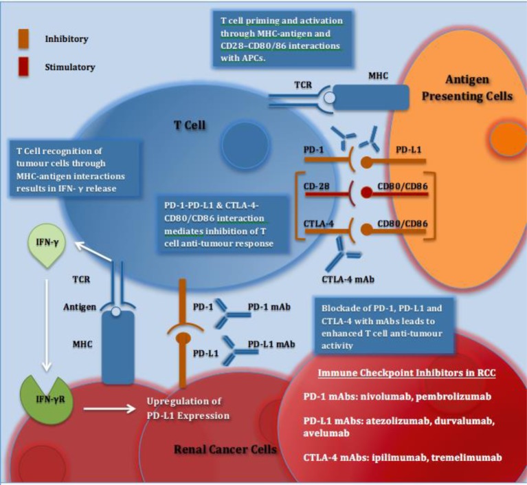
Figure 1. Immune checkpoints and immune checkpoint inhibitors in RCC.
Recognition of tumour cells and APCs via MHC–antigen interactions with TCRs activates T cells. IFN-γ released from T cells results in up-regulation of PD-L1 expression. PD-1 is expressed on activated T cells and on interaction with PD-L1 on tumour cells or APCs results in inhibition of T cell antitumour response. CTLA-4 is expressed on T cells and on interaction with its ligands CD80/CD86 on APCs, T-cell proliferation and T-cell effector function is reduced. CD28 is a co-stimulatory T-cell molecule, which has a lower affinity than CTLA-4 for their shared ligands; CD80/CD86. Blockade of PD-1, PD-L1 and CTLA-4 with mAbs stimulates an enhanced antitumour response and has shown efficacy in aRCC. Abbreviations: aRCC, advanced renal cell cancer; APC, antigen presenting cell; CD28, cluster of differentiation 28; CD80, cluster of differentiation 80; CD86, cluster of differentiation 86; CTLA-4, cytotoxic T lymphocyte associated protein 4; IFN-γ, interferon-γ; IFN-γR, interferon-γ receptor; mAb, monoclonal antibody; PD-1, programmed death-1; PD-L1, programmed death ligand 1; TCR, T-cell receptor.

