. 2017 Oct 23;9(1):52–61. doi: 10.1039/c7sc03939e

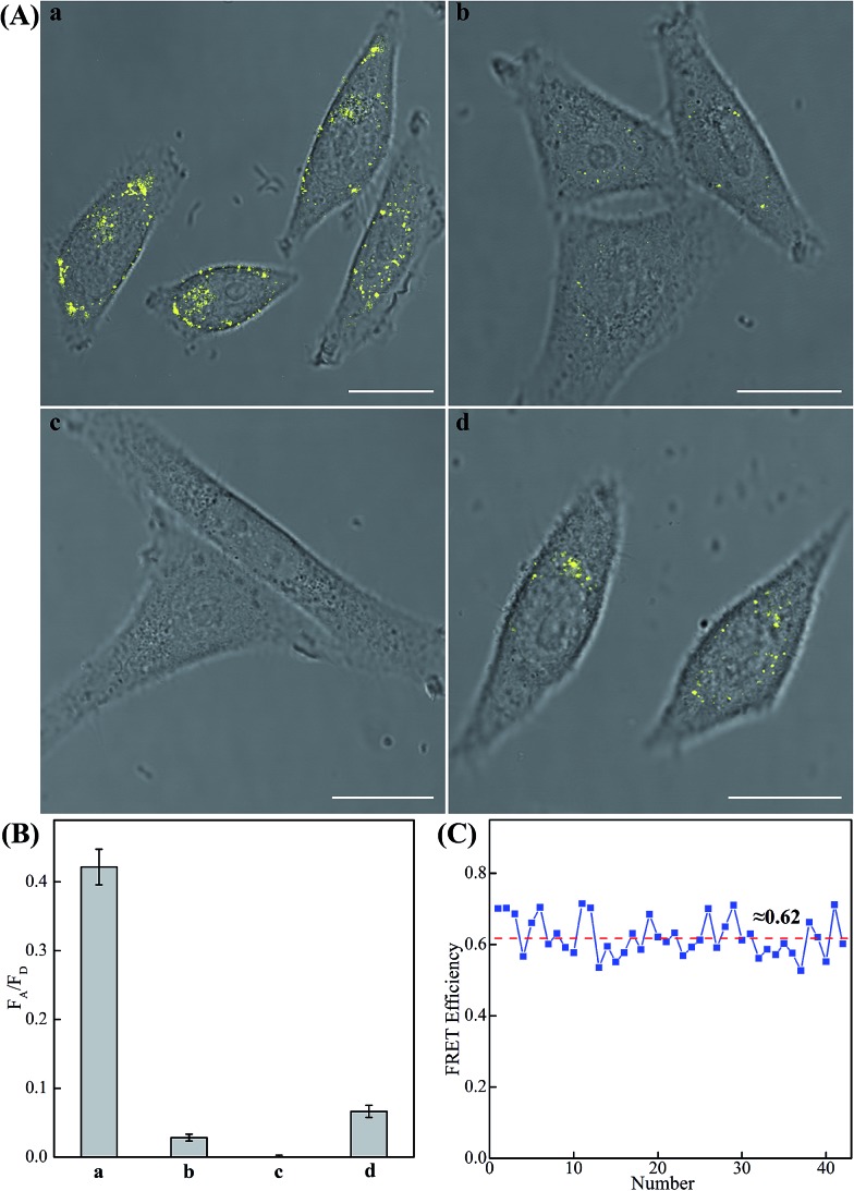
Fig. 6. (A) Living cell analysis of miR-21 based on C-HCR or HCR strategy and FRET transduction (in the form of FA/FD). Confocal laser scanning microscopy (CLSM) imaging of miR-21 in (a) routine MCF-7 live cells by C-HCR amplifier, (b) routine MCF-7 live cells by conventional HCR amplifier (H6-excluded C-HCR), (c) MCF-7 live cells treated with a chemically modified miR-21 inhibitor by C-HCR amplifier, and (d) HeLa live cells by C-HCR amplifier. All of the aforementioned living cells were transfected and incubated with miR-21-targeting C-HCR mixture at 37 °C for 2 h. All scale bars correspond to 20 μm. (B) Statistical histogram analysis of the relative fluorescence intensity (FA/FD) of the above four cell samples through C-HCR imaging system. (C) Determination of the FRET efficiency of the C-HCR-imaging system collected from a large number of MCF-7 living cells.

Fig. 6