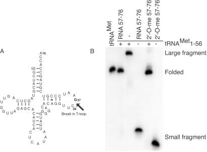
Figure 1.
Folding of fragmented E. coli tRNAMet. (A) Secondary structure of tRNAMet with a break between positions 56 and 57 in the phosphodiester backbone is indicated by the arrow. (B) Folding of fragmented tRNAMet is monitored with nondenaturing PAGE. Lanes: tRNAMet, E. coli tRNAMet; RNA 57–76, unsubstituted smaller RNA fragment; 2′-O-me 57–76, fully 2′-O-methyl substituted smaller fragment. (+) and (−) indicate presence and absence, respectively, of the 5′ large fragment (positions 1–56). Mobility of the folded tRNAMet, 5′ large fragment, and 3′ small fragment are indicated on the right.

