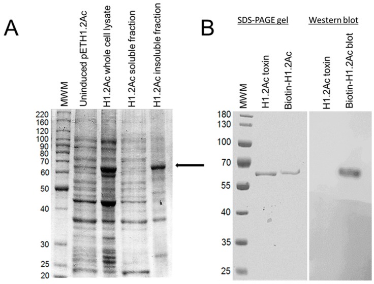
Figure 2.
Expression, purification, and biotinylation of H1.2Ac. The arrow indicates the expected size for the H1.2Ac protoxin. (A) SDS-PAGE gel showing proteins expressed by transformed E. coli cultures with or without IPTG induction, as stated in the figure. The H1.2Ac protein was only detected in the insoluble fraction of the total cell lysate. (B) Electrophoretic detection of purified trypsin-activated H1.2Ac and Western blot detecting biotinylated H1.2Ac using enhanced chemiluminescence. MWM, molecular weight markers in kDa.

