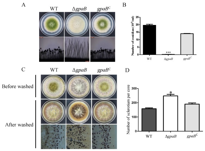
Figure 4.
gpaB is involved in conidiation and sclerotia formation in A. flavus. (A) Colony and conidiophore morphology among the WT, ΔgpaB and gpaBC were observed after being grown on PDA agar medium for five days in the dark; (B) The number of conidia of the indicated strains was measured after being grown on PDA agar for five days; (C) Sclerotia formation of the indicated strains grown on sclerotia-inducing Wickerham (WKM) medium was detected. To visualize the sclerotia, 75% ethanol was sprayed on the WKM plates to remove the conidia; (D) The number of sclerotial was counted as in (C). Error bars represent the standard deviation from four replicates, and asterisks, “***” or “*”, represent significant differences compared to the wildtype according to the t-test with p < 0.001 and p < 0.05, respectively. The experiments were conducted with four replicates for the indicated strain and were repeated three times.

