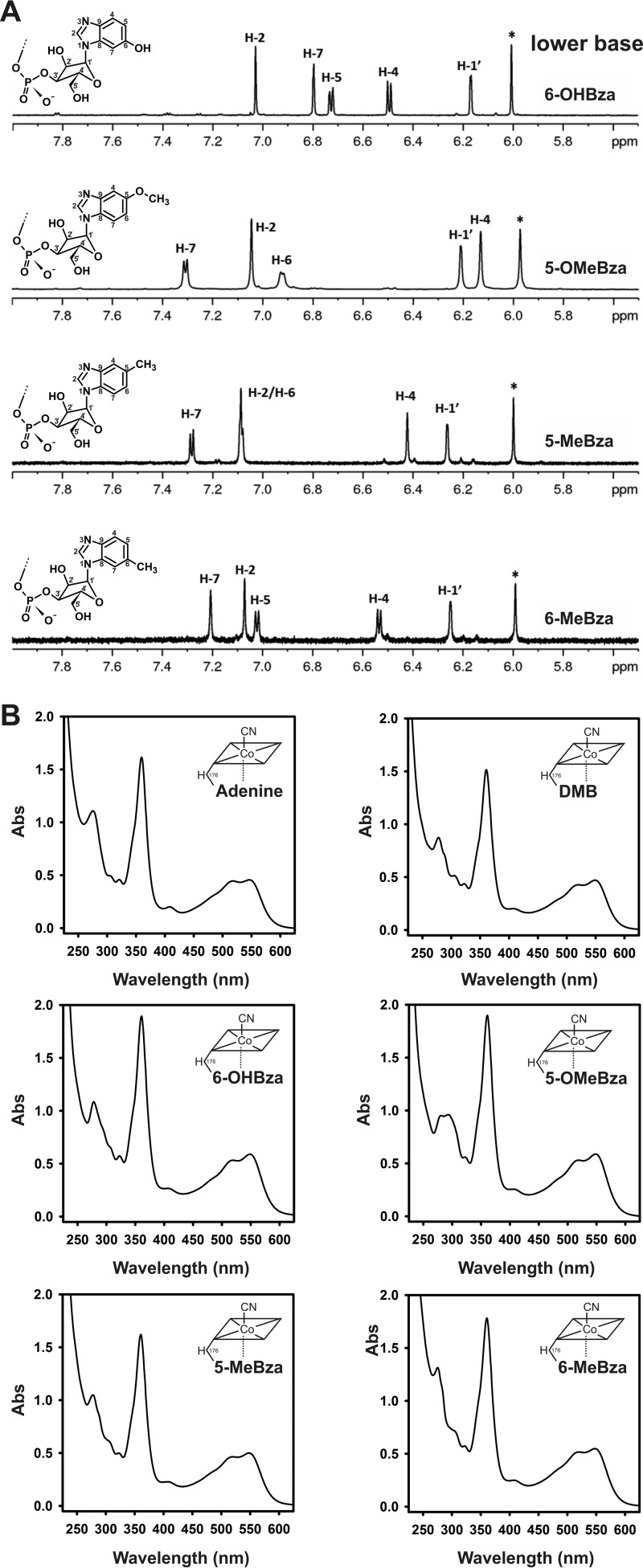
FIG 2.
Characterization of the isolated NCbas. (A) Low field range of 1H-NMR spectra of 6-OHBza-NCba, 5-OMeBza-NCba, 5-MeBza-NCba (peak 1 in Fig. 1C), and 6-MeBza-NCba (peak 2 in Fig. 1C). The depicted sections show the signals for the respective benzimidazolyl moieties and the signal for the anomeric position of the α-ribosyl unit. The signal marked with an asterisk belongs to position 10 of the corrin scaffold. (B) UV/Vis-absorbance spectra of the newly purified NCbas in comparison to those of the authentic adeninyl-NCba (norpseudovitamin B12) and the DMB-NCba (norvitamin B12).

