FIG 3.
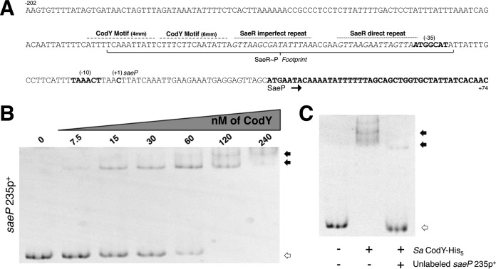
S. aureus CodY interacts with the sae P1 upstream region. (A) The region surrounding the sae P1 regulatory region is displayed (coding strand; 5′ to 3′). The known translated sequence, transcriptional start site (+1), −10, and −35 sites are displayed and annotated in boldface. The identified SaeR∼P footprint is bracketed with the direct and indirect repeat motifs indicated above the sequence with a dotted line. A dashed line above the sequence identifies the putative CodY motifs. mm, mismatch. (B) Purified SaCodY-His6 protein was incubated with a 6-FAM-labeled 235-bp DNA fragment containing the upstream regulatory region of saeP in the presence of ILV and GTP. The unbound fragment is indicated with open arrows while CodY-DNA complexes are indicated by filled arrows. Increasing amounts of CodY monomer were incubated with the fragment. The molar concentration (of monomeric protein) used in each reaction mixture is indicated above each lane. (C) CodY monomer (120 nM) was incubated with the fragment in the presence of a 30× molar excess of unlabeled probe. The presence of each protein and unlabeled probe is indicated beneath each lane.

