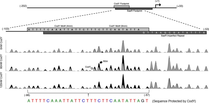
FIG 4.
DNase I footprinting reveals that the CodY and SaeR binding sites overlap. A 5′-FAM-labeled saeP235p+ fragment incubated in the presence of 0, 60, and 120 nM purified CodY or bovine serum albumin (BSA; control) was challenged with 0.075 to 0.1 U of DNase I. The resulting fragments were separated using capillary electrophoresis and aligned to the sequenced PCR product as a reference. Relative fluorescence (y axis) is displayed as a function of nucleotide position (x axis). Reaction mixtures containing CodY (gray trace) were compared to control reaction mixtures containing bovine serum albumin (black trace). A drop in peak intensity at a given nucleotide position indicates protection by CodY. CodY binds to a 31-bp region (colored nucleotides). Data are representative of at least two independent experiments.

