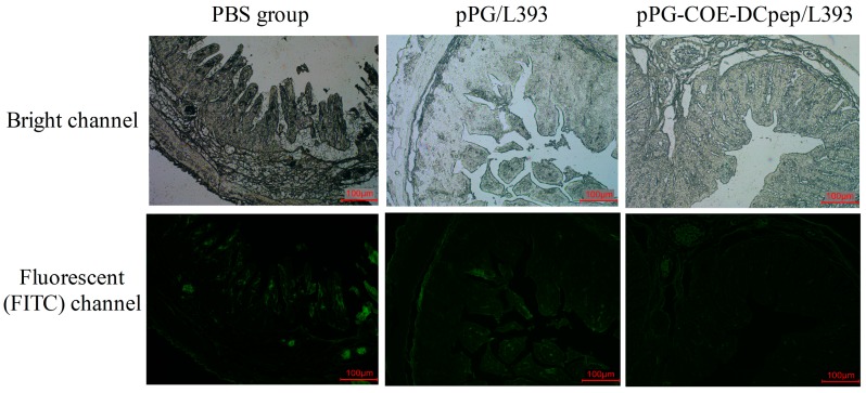
Figure 6.
PEDV distribution in the jejunum. The piglets were orally infection with PEDV (1 × 10⁶ PFU/mL) via oral-gastric gavage. The PEDV distribution patterns were detected in the villous by an immunofluorescence assay using rabbit polyclonal antibody against PEDV. The top pictures are from the light channel, and the bottom pictures are from the fluorescent (FITC) channel. The upper and lower panel were from the same section. The pictures are representative of sections derived from each group.

