Figure 2.
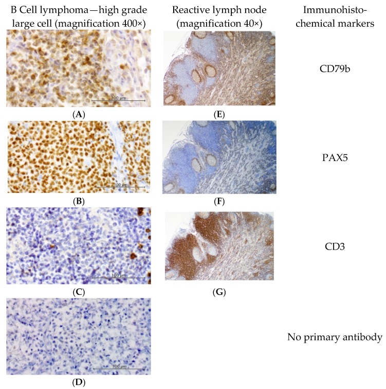
Immunohistochemical labelling. Results from a representative B cell lymphoma (left) demonstrate strong membranous labelling of proliferating cells with (A) anti-CD79b B-cell marker (1:5000), and nuclear labelling with (B) anti-PAX5 B-cell marker (1:100). (C) Anti-CD3 T-cell marker showed strong membranous staining of a few scattered small lymphocytes (1:400). Omission of the primary antibody (D) shows minimal background staining. The positive tissue control (E–G, reactive feline lymph node, right) demonstrates appropriate staining of B and T cells. Slides counterstained with Whitlock’s hematoxylin. Positive control sections shown at 40× magnification; all other images are 400× magnification.

