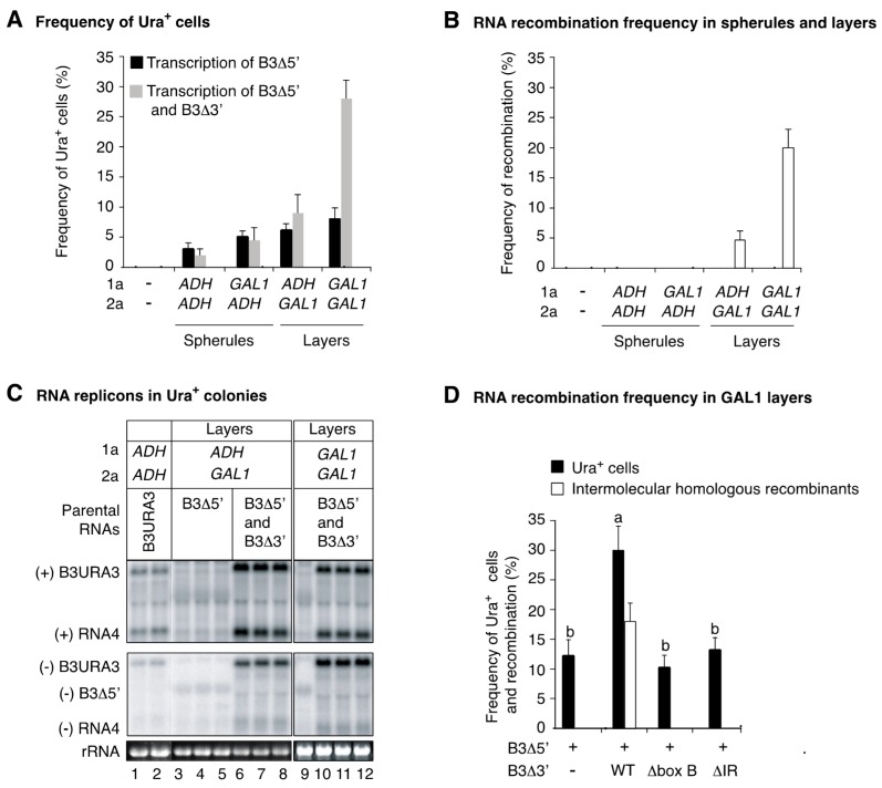
Figure 8.
Intermolecular RNA recombination in layers after one yeast generation. Cells were incubated for 15 h in liquid media (8 mL) containing galactose and 500 μM CuSO4. 1a and 2apol were expressed from the ADH or GAL1 promoters to induce the formation of spherules or layers. Ura+ cells were identified by uracil selection on plates. The average sample size was 700 cells per treatment. (A) Frequency of Ura+ cells after transcription of B3Δ5′ alone or in combination with B3Δ3′. Transcription of B3Δ5′ alone supported Ura+ colony formation by directing RNA4 transcription. A significant (p < 0.01) increment in the number of Ura+ cells was obtained in layer-forming conditions. (B) Frequency of intermolecular RNA recombination in Ura+ cells described in (A). (C) The presence of intermolecular RNA recombinants was confirmed by analyzing 48 Ura+ colonies individually grown for 36 h in cultures (8 mL) lacking uracil and copper. RNA was extracted an analyzed by Northern blotting using URA3 probes. Representative blots are shown. B3URA3 was a size marker (lanes 1 and 2). (+) and (−) indicate RNA of positive or negative polarity, respectively. As illustrated in Figure 7A, B3Δ5′ supported negative-strand B3Δ5′ synthesis and RNA4 transcription (lanes 3, 4, 5 and 9). Lanes 6, 7, 8, 10, 11 and 12 are intermolecular RNA recombinants. (D) Frequency of Ura+ cells obtained after transient transcription of B3Δ5′ and wild type or mutant (box B or intergenic region deletion) B3Δ3′ for one yeast generation in GAL1 layers. Induction of transcription, selection, and identification on intermolecular RNA recombinants was as in (A,B). Bars represent the average and standard error of three biological replicates. Bars with the same letter are not significantly different (Tukey's test with alpha = 0.01).

