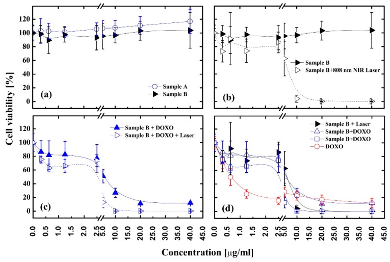
Figure 7.
Cell viabilities of HepG2 cells after being incubated with different nanomaterials at various concentrations. (a) Sample A—Fe3O4@PDA and Sample B—Fe3O4@PDA@SH-βCD; (b) Sample B—with and without laser irradiation; (c)Sample B—loaded with DOXO with and without laser irradiation; (d) Replotted data from (b,c) plus free DOXO as the reference.

