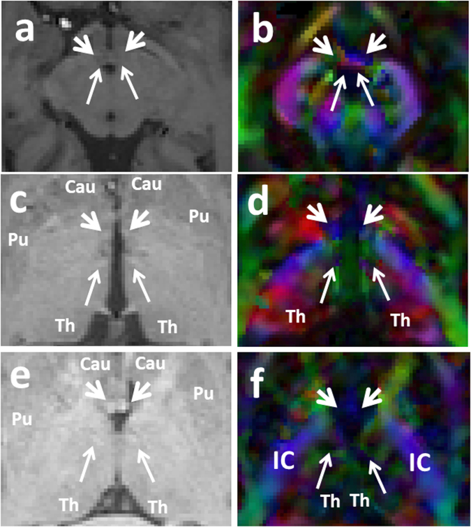
Figure 3.
Three successive color-coded DTI map and corresponding T1 weighted MRI sequences in axial planes from caudal to cranial demonstrating the relationship of the postcommissural fibers of the fornix and stria terminalis (pointed by short arrows), with the MTT (pointed by long arrows) as they course parallel to each other. Four bright spots are visible on the T1 weighted images at the level of mammillary bodies and higher levels (a,c,e). The two frontal bright spots represent the forniceal columns. The two dorsal bright spots represent the MTT on both sides (Fig. a,c,e). As it’s visible on Fig. 3a,b, the ascending fibers of the MTT arise from the mammillary bodies (MB), course posteriorly and then cranially behind the poscommissural fibers of the fornix and stria terminalis (pointed by short arrows). The MTT then rises within the anterior thalami (Th) projecting more laterally as its visible on the (e and f). The MTT fibers then terminate in the anterior and medial thalamic nuclei in each side. Cau = caudate head; IC = internal capsule; Pu = putamen; Th = thalamus.

