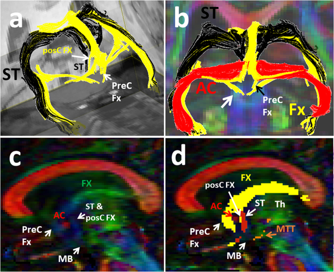
Figure 4.
Three-dimensional reconstructions of the fornix (in yellow) and the stria terminalis (in black, a and b). The vertical blue fibers anterior to the anterior commissure (AC) shown in c, are the precommisural (PreC) fibers of the fornix (Fx). The vertical blue fibers posterior to the anterior commissure (AC) shown in c, are a combination of the postcommissural (PosC) fibers of the fornix and stria terminalis (ST). The corresponding trajectory of the fornix (yellow) and stria terminalis (shown in red) are shown in d. The precommissural (PreC) and postcommissural (PosC) fibers of the fornix are shown in yellow color anterior and posterior to the AC respectively. The trajectory of the MTT is shown (in orange in d) arising from the mammillary bodies (MB) and coursing posteriorly and then cranially toward the thalamus (Th).

