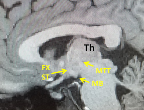
Figure 5.

Sagittal T1 weighted MRI showing the trajectory of the MTT in relation with the precommissural fibers of the fornix and stria terminalis (Fx, ST). The MTT is visible by a T1 hypointense tract seen arising from the mammillary body (MB), projecting posteriorly and then cranially within the thalamus. The superior aspect of the MTT is not visible in this sagittal plane since the MTT projects more laterally as it ascends within the thalamus. The postcommissural fibers of the fornix and stria terminalis are visible by a T1 hypointense bundle vertically arising from the mammillary bodies.
