Abstract
Basal septal hypertrophy is a rare and unique anatomical finding associated with hypertrophic cardiomyopathy (HCM). It is also described as a sigmoid hypertrophy and is linked with aging and chronic hypertension. Takotsubo cardiomyopathy is a transient cardiomyopathy that occurs during periods of high physical or emotional stress. Its occurrence with HCM is relatively common; however, this presentation occurs more often with the classic asymmetrical septal hypertrophy or the apical variant. This case demonstrates its coexistence with isolated sigmoid hypertrophy in an elderly, hypertensive female with severe ischemic bowel disease.
Key Words: Basal hypertrophy, bowel ischemia, takotsubo
INTRODUCTION
Basal septal hypertrophy is a unique morphological finding in patients with hypertrophic cardiomyopathy (HCM). This condition is also called sigmoid hypertrophy and is associated with aging and chronic hypertension.[1] Takotsubo is a transient cardiomyopathy that occurs during periods of high physical or emotional stress. It typically presents with substernal chest pain, electrocardiogram changes which include ST elevation, ST depression, T-wave inversion, pathologic Q-waves and QT prolongation, shortness of breath, or cardiogenic shock.[2,3] This disease entity can clinically manifest in patients with HCM but typically occurs more often with classic asymmetrical septal hypertrophy or the apical variant.[1,2,3] Our interesting case demonstrates this unique coexistence, and its recognition is critical for the management and treatment, especially in the setting of cardiogenic shock, as inotropic agents are likely to aggravate and worsen the clinical condition.
CASE REPORT
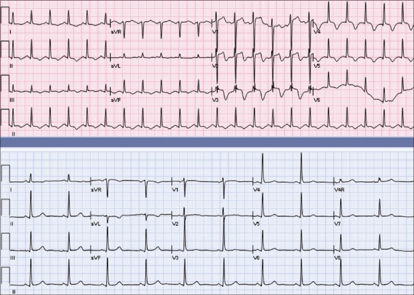
A 74-year-old female with a past medical history of hypertension and hyperlipidemia presented to the ER with chest pain. 7-months ago she had a sigmoid resection secondary to diverticulitis. She was hypotensive, tachycardic, and in respiratory distress. On physical examination, she had bibasilar rales, peripheral edema, and jugular venous distension. Her blood pressure was 94/60, and heart rate was 133 and hypoxic. She was started on intravenous furosemide and metoprolol and placed on noninvasive mechanical ventilation with limited improvement. Chest X-ray demonstrates bilateral pleural effusions. Electrocardiogram revealed T-wave inversions of the inferior leads with ST elevations in V1–V3 [Figure 1]. Troponin was elevated at 2.43, CPK 514, CK-MB 229, and BNP >5000 with lactic acid 2.1. Echo [Figures 2 and 3] demonstrates hypokinesis from the mid-anterior wall to the apex with systolic anterior motion (SAM) of the mitral valve and significant mitral regurgitation (MR). Systolic function was decreased with ejection fraction (EF) 44%. She was administered dual-antiplatelet therapy and sent for emergent catheterization due to suspected anterior wall infarction. Cardiac catheterization [Figure 4] demonstrated nonobstructive coronary artery disease of the left anterior descending (LAD) with an EF of 30%. There was also an increased left ventricular outflow tract (LVOT) pressure gradient [Figure 5] secondary to SAM. These findings are suggestive of Takotsubo with mid-LV obstruction. Cardiac output improved with the use of phenylephrine and beta-blockers, but the patient's condition continued to deteriorate over the next 3 days. She became febrile with leukocytosis, lactic acid increased, and blood cultures drawn became positive for Candida glabrata. The patient became septic with peritoneal signs, at which time she is taken back to the operating room (OR). Two areas of infarcted small bowel are discovered and subsequently resected up to the cecum. Over the course of the next 3 weeks, the patient was stabilized off vasopressors. Repeat echo demonstrates EF of 65% with the resolution of apical ballooning and wall abnormalities [Figure 6]. However, LVOT turbulence with gradient >30 mmHg was remarkable with sigmoid hypertrophy. Severe MR with posterior jet was also persistent, due to malcoaptation of the leaflets. Aortic insufficiency was also evident. Clinically, the patient's clinical condition improved during hospital stay.
Figure 1.
EKG showing ST-segment elevation on anteroseptal leads with reciprocal changes on inferolateral leads
Figure 2.

Two-chamber view with color Doppler showing severe eccentric mitral regurgitation Jet
Figure 3.

Two-chamber view showing hypokinesis from mid anterior wall to apex
Figure 4.

Left ventriculogram showing ventricular apical ballooning
Figure 5.

Pressure tracing during pull back showing the increased LVOT gradient
Figure 6.

Chamber View showing resolution of apical ballooning and wall motion abnormalities
DISCUSSION
Takotsubo cardiomyopathy (TCM), also known as stress cardiomyopathy, transient apical ballooning, or broken heart syndrome, is described by Armstrong et al., as the shape of the LV being similar to a Japanese octopus trap, with a round bottom and narrow neck.[2]
Cardiac catheterization was the initial intervention in our patient since she presented with myocardial hypokinesis, elevated troponins, and ST changes. Infarction of the LAD is a rare cause of LVOT obstruction with SAM, and hence, had to be ruled out initially.[3] Basal hyperkinesis results in increased velocity through the LVOT, and this can, in turn, result in anterior displacement of the mitral valve leaflet into the septum through the Venturi effect.[4] As was the case in our patient, this disease entity is most common in older women. Similar findings may be found in TCM and neurogenic myocardial stunning, which typically occur after intracerebral hemorrhage.[3] In our patient, with nonobstructive disease of the LAD, TCM with coexisting HCM physiology is the most likely diagnosis.
The presence of SAM is highly suggestive of LVOT obstruction, which is known to induce MR.[3,4] TCM has been associated with both inducing LVOT obstruction and masking existing HCM pressure gradients. In our patient, initial echocardiographic findings demonstrated apical ballooning with reduced EF, SAM, and MR. Three weeks later, there was resolution of TCM, but the obstruction with LVOT gradient >30 mmHg persisted.
The “sigmoid hypertrophy” which was identified is related to aging, but her intraventricular septum and posterior wall were not hypertrophied. There was also significant aortic regurgitation present. Sigmoid morphology is isolated hypertrophy of the basal anteroseptum, which causes a type of subaortic stenosis.[5] Aortic regurgitation develops from chronic turbulence across the valve.[6] Basal hypertrophy has been linked to long-standing hypertension, which was noted in our patient.[1] It is likely that Takotsubo developed in the setting of infection and surgery, and this, in turn, resulted in a decrease in the flow out of an already narrowed outflow tract. Conversely, sigmoid hypertrophy with catecholamine excess and hypovolemia contributes to a stress-induced cardiomyopathy.
There are earlier studies that suggest that LVOT obstruction might be present in 25% of patients with TCM. Echocardiography reveals a typical septal bulge associated with SAM of the mitral valve and MR similar to the findings associated with HOCM.[7] Mid-ventricular septal thickening is a structural abnormality associated with LVOT obstruction, and this finding could potentially cause severe, transient LV mid-cavity obstruction in the presence of increased catecholamine levels.[8] It is still unclear if LVOT obstruction is a result or cause of stress cardiomyopathy.[9]
The earlier similar case report all report, on clinical presentation, a pressure gradient between the aorta and apical LV. There are two possible explanations for this clinical presentation. During the acute attack, there might be severe systolic myocardial dysfunction with subsequent low cardiac output which masks the gradient. This picture is similar to the scenario of patients with severe aortic stenosis with low aortic valve pressure gradients due to left ventricular systolic dysfunction. Another explanation might include basal septal involvement in the pathophysiology of TCM resulting in a transient pseudonormalization of the systolic pressure gradient.[10]
Dynamic outflow obstruction is often exacerbated in the setting of hypovolemia with the use of inotropic agents; thus, adrenergic drugs are discouraged in patients with HCM physiology. An intra-aortic balloon pump, an alternative or adjunct to inotropic drugs, is also discouraged in these situations because the increase in afterload will increase the obstruction. Phenylephrine was our choice for maintaining mean arterial pressure >65 during the period of stress cardiomyopathy because it is an alpha-specific agonist that increases afterload. It does not affect chronotropy; thus it is a good choice to facilitate diastolic filling, especially in the setting of tachycardia. Management of HCM in cardiogenic shock is precarious. Additional supportive care with fluids and cautious use of beta-blockers are recommended.[7,8,11] Patients who have sustained systolic dysfunction in the setting of HCM may be considered for implantable cardioverter defibrillator therapy.
CONCLUSION
TCM is usually a benign reversible condition. However, in the clinical setting of basal hypertrophy causing LVOT obstruction and cardiogenic shock, it can potentially be severe and fatal. Cardiologists, intensivisits, and clinicians alike need to recognize and comprehend the pathophysiology behind this unique clinical manifestation so that they may adjust their management and treatment accordingly.
Declaration of patient consent
The authors certify that they have obtained all appropriate patient consent forms. In the form the patient(s) has/have given his/her/their consent for his/her/their images and other clinical information to be reported in the journal. The patients understand that their names and initials will not be published and due efforts will be made to conceal their identity, but anonymity cannot be guaranteed.
Financial support and sponsorship
Nil.
Conflicts of interest
There are no conflicts of interest.
REFERENCES
- 1.Kelshiker MA, Mayet J, Unsworth B, Okonko DO. Basal septal hypertrophy. Curr Cardiol Rev. 2013;9:325–30. doi: 10.2174/1573403X09666131202125424. [DOI] [PMC free article] [PubMed] [Google Scholar]
- 2.Armstrong WF, Ryan T, Feigenbaum H. Feigenbaum's Echocardiography. 7th ed. Philadelphia: Wolters Kluwer Health/Lippincott Williams & Wilkins; 2010. [Google Scholar]
- 3.Lee JW, Kim JY. Stress-induced cardiomyopathy: The role of echocardiography. J Cardiovasc Ultrasound. 2011;19:7–12. doi: 10.4250/jcu.2011.19.1.7. [DOI] [PMC free article] [PubMed] [Google Scholar]
- 4.Turer AT, Samad Z, Valente AM, Parker MA, Hayes B, Kim RJ, et al. Anatomic and clinical correlates of septal morphology in hypertrophic cardiomyopathy. Eur J Echocardiogr. 2011;12:131–9. doi: 10.1093/ejechocard/jeq163. [DOI] [PubMed] [Google Scholar]
- 5.Aboulhosn J, Child JS. Left ventricular outflow obstruction: Subaortic stenosis, bicuspid aortic valve, supravalvar aortic stenosis, and coarctation of the aorta. Circulation. 2006;114:2412–22. doi: 10.1161/CIRCULATIONAHA.105.592089. [DOI] [PubMed] [Google Scholar]
- 6.Fifer MA, Vlahakes GJ. Management of symptoms in hypertrophic cardiomyopathy. Circulation. 2008;117:429–39. doi: 10.1161/CIRCULATIONAHA.107.694158. [DOI] [PubMed] [Google Scholar]
- 7.Nef HM, Möllmann H, Akashi YJ, Hamm CW. Mechanisms of stress (Takotsubo) cardiomyopathy. Nat Rev Cardiol. 2010;7:187–93. doi: 10.1038/nrcardio.2010.16. [DOI] [PubMed] [Google Scholar]
- 8.Merli E, Sutcliffe S, Gori M, Sutherland GG. Tako-Tsubo cardiomyopathy: New insights into the possible underlying pathophysiology. Eur J Echocardiogr. 2006;7:53–61. doi: 10.1016/j.euje.2005.08.003. [DOI] [PubMed] [Google Scholar]
- 9.Afonso L, Bachour K, Awad K, Sandidge G. Takotsubo cardiomyopathy: Pathogenetic insights and myocardial perfusion kinetics using myocardial contrast echocardiography. Eur J Echocardiogr. 2008;9:849–54. doi: 10.1093/ejechocard/jen192. [DOI] [PubMed] [Google Scholar]
- 10.Daralammori Y, El Garhy M, Gayed MR, Farah A, Lauer B, Secknus MA, et al. Hypertrophic obstructive cardiomyopathy masked by Takotsubo syndrome: A case report. Case Rep Cardiol. 2012;2012:486427. doi: 10.1155/2012/486427. [DOI] [PMC free article] [PubMed] [Google Scholar]
- 11.El Mahmoud R, Mansencal N, Pilliére R, Leyer F, Abbou N, Michaud P, et al. Prevalence and characteristics of left ventricular outflow tract obstruction in Tako-Tsubo syndrome. Am Heart J. 2008;156:543–8. doi: 10.1016/j.ahj.2008.05.002. [DOI] [PubMed] [Google Scholar]


