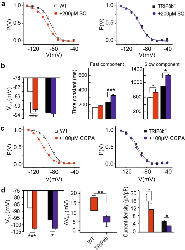
Fig. 4.
Modulation of Ih in TC neurons by cAMP. a Graphs showing the mean steady-state activation curves of I h in thalamocortical (TC) neurons of WT (n = 8/7 cells) and TRIP8b−/− (n = 11/8 cells) mice. Slices were incubated either with 200 µM (dots) or without (squares) the adenylyl cyclase inhibitor SQ22536. Incubation with SQ22536 shifted the voltage-dependent activation (V 0.5) of I h to more hyperpolarizing potentials without having any effects on I h in TRIP8b−/− TC neurons (left panel, ANOVA followed by Student’s t tests, *** indicates p < 0.001). b (middle and right panel) The activation kinetics of Ih in both TRIP8b−/− and WT TC neurons were slowed down in SQ22536 treated cells. c Graphs showing the mean steady-state activation curves of I h in WT (n = 5 cells) and TRIP8b−/− (n = 6 cells) TC neurons of the VB complex following bath application of 100 µM CCPA (dots). d Although CCPA shifted the voltage-dependent activation of I h to negative potentials in both groups (left panel), the hyperpolarizing shift in V 0.5 of I h (ΔV 0.5, middle panel) in thalamic relay neurons of TRIP8b−/− mice was significantly smaller compared to WT TC neurons. The reduction in I h current density after application of CCPA in both groups is depicted in the bar graph (right panel). Repeated-measures ANOVA followed by Student’s t tests, *, **, *** indicate p < 0.05, p < 0.01, and p < 0.001, respectively

