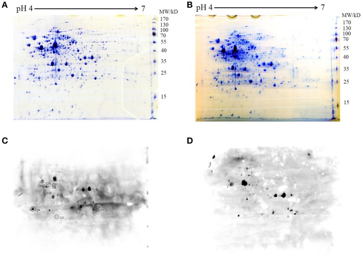
Figure 2.
Two-dimensional map of the whole proteins of (A) WT and (B) Δstk. The gels were stained with Coomassie blue (A,B) or electroblotted and probed with an anti-pThr antibody (C,D). Protein spots C11 to C36 corresponding to phosphorylated proteins were excised and analyzed by MALDI-FTMS. Molecular weights are indicated on the right.

