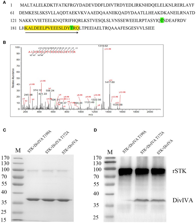
Figure 4.
Identification of DivIVA phosphorylated sites. (A) Protein sequence of DivIVA: The peptide containing a phosphorylated signal is highlighted in yellow and labeled with arrows. The threonine residues introduced by mutagenesis are labeled in green. (B) Mass spectra showing that DivIVA is phosphorylated at threonine 199. Recombinant DivIVA was incubated with rSTK and then subjected to trypsin digestion. (C) rSTK incubated with mutant DivIVA proteins were separated by SDS-PAGE and then stained with Coomassie blue. (D) An anti-pThr antibody was used to assess the effect of mutant DivIVA proteins.

