Figure 7.
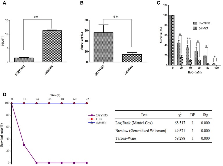
Virulence attenuation of the ΔdivIVA mutant strain. (A) Evaluation the anti-phagocytotic ability of S. suis strains in macrophage RAW264.7 cells. (B) The ΔdivIVA strain showing decreased resistance to phagocytosis and killing by neutrophils. The 106 CFU of the wild-type and mutant strains was incubated with PMNs at a bacteria-to-cell ratio of 10:1. The cells were lysed after 1 h of incubation, and the survival percentage of each strain was calculated as follows: (CFUPMN+/CFUPMN) × 100%. The data are expressed as the mean and standard deviation of three independent experiments (**P < 0.01). (C) H2O2 survival test of the WT and the ΔdivIVA mutant. S. suis 2 cultures at the mid-exponential phase (106 CFU) were incubated with increasing concentrations of H2O2 at room temperature for 15 min, and viable counts were monitored. The assay was performed in triplicate. A statistically significant killing effect of H2O2 on the WT05ZYH33 and ΔdivIVA mutant was observed (*P < 0.05; **P < 0.01). (D) Survival curves of mice infected with S. suis 2 strains. Four-week-old BALB/c mice were challenged intraperitoneally with 108 CFU bacteria, and the survival time was monitored. Kaplan-Meier survival analysis was performed with three different tests to evaluate the significant difference of the survival rates among the three groups.

