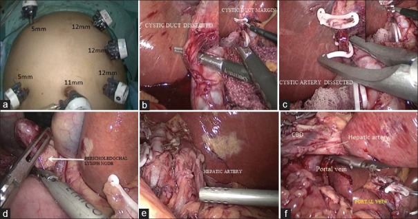
Figure 2.
(a) Port placement; (b) cystic duct ligation with inset showing excision cystic duct margin; (c) cystic artery ligation and division; (d) showing excision of pericholedochal lymph node; (e) hepatic artery after lymph node excision; (f) portal vein after lymph node excision with inset showing contralateral side of portal vein

