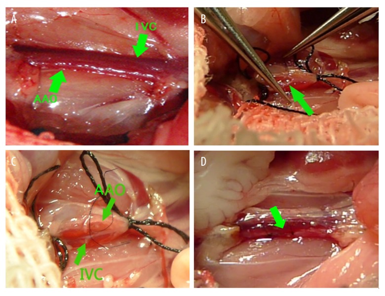
Figure 1.
Surgical procedure of creation of the shunt. (A) The abdominal aorta (AAo) and inferior vena cava (IVC) were tightly linked together; (B) Blood in the abdominal aorta and the inferior vena cava were blocked and a transverse incision were made in the abdominal aorta’s anterior wall to expose the contralateral wall, and then the wall were cut by micro-scissors; (C) Normal saline was injected from the abdominal aorta and the inferior vena cava was filled with normal saline; (D) After the shunt was made, the inferior vena cava pulsed with arterial blood.

