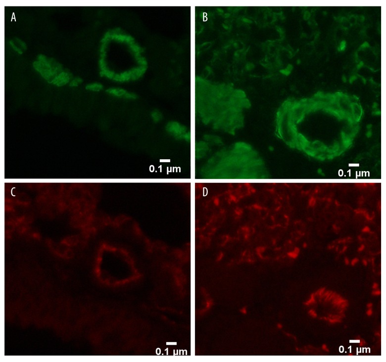
Figure 5.
Pulmonary vascular remodeling in the 12th week. (A) Pulmonary vascular remodeling in the 12th week (×400) showing proliferation of smooth muscle cells (stained in green) in the small shunt group; (B) Pulmonary vascular remodeling in the 12th week (×400) showing obvious proliferation of smooth muscle cells (stained in green) in the heavy shunt group; (C) Pulmonary vascular remodeling in the 12th week (×400) showing a monolayer of endothelial cells (stained in red) in the small shunt group; and (D) Pulmonary vascular remodeling in the 12th week (×400) showing proliferation of endothelial cells (stained in red) in the heavy shunt group.

