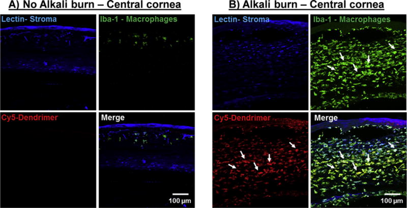
Fig. 4. Biodistribution of subconjunctivally injected dendrimers.

Fluorescently labelled dendrimers (D-Cy5) in gel formulations were injected subconjuntivally and the biodistribution was assessed 7 days after injection. Corneal stroma (Blue, Lectin), Macrophages (Green, Iba-1), Dendrimer (Red, Cy5). A) A central cross section of a normal cornea with regular tissue architecture; very few corneal Iba-1 stained cells (macrophages) are present; dendrimers are not co-localized in the macrophages. B) An alkali burnt central cornea infiltrated with Iba-1 positive cells (macrophages). Cy5 signals (dendrimer) are co-localized in the Iba-1 stained cells demonstrating dendrimer’s intrinsic targeting capability towards inflammation. Scale bar 100 μm. (For interpretation of the references to colour in this figure legend, the reader is referred to the web version of this article.)
