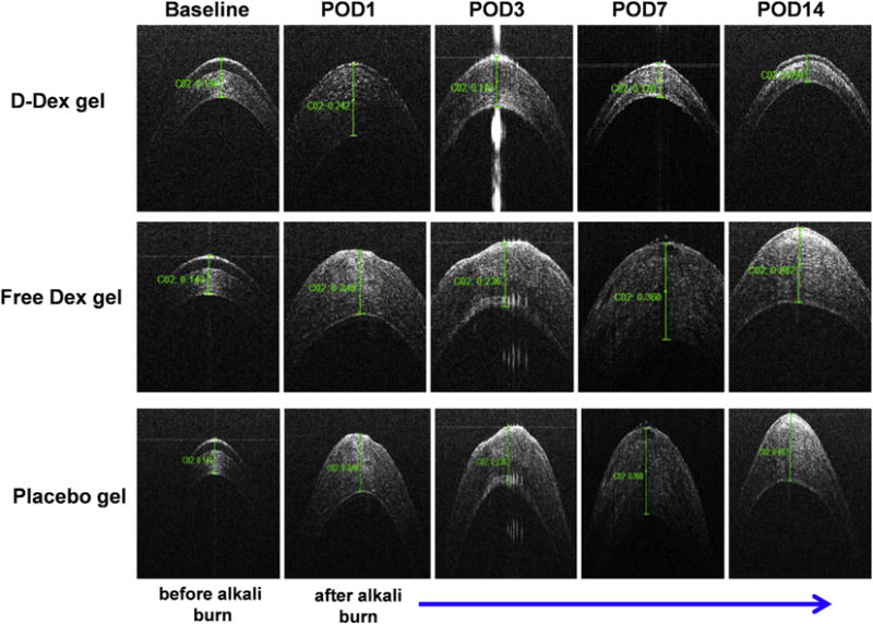
Fig. 5. Anterior segment optical coherence tomography (OCT) imaging of the cornea for assessment of central corneal thickness (CCT).

Top panel: OCT images of the central cornea of a D-Dex gel treated eye demonstrate near-normal corneal architecture at POD 7 and 14 when compared to its baseline. These images suggest that inflammation has subsided. Middle panel: OCT images of a central cornea treated with free-Dex gel demonstrate a thin irregular epithelial layer and stromal edema which suggest ongoing inflammation. Bottom panel: OCT images of a central cornea treated with placebo gel has similar characteristics as the free-Dex treated eye.
