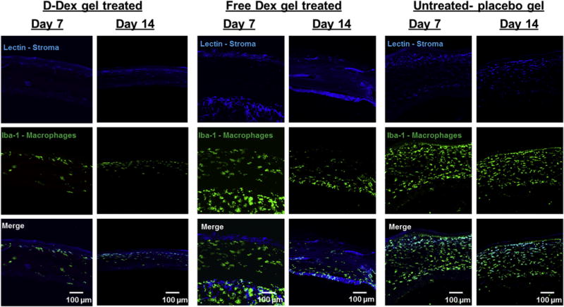
Fig. 8. Confocal microscope images of corneal cross sections.

Left panel: a minimal amount of Iba-1 stained cellular infiltrate (macrophages) is observed at post-operative day 7 and 14. Middle pane and right panels: Unlike the D-Dex gel group, both free-Dex gel and placebo gel groups have a persistent IBA-1 stained cellular infiltrate (macrophages) at post-operative days 7 and 14. Scale bar 100 μm.
