. 2017 Nov 16;9(4):841–849. doi: 10.1039/c7sc03712k

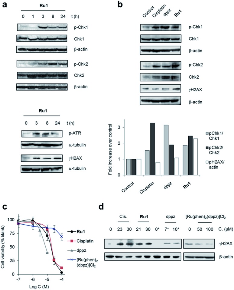
Fig. 2. (a) DNA damage response (DDR) activation by Ru1. Whole-cell extracts of OE21 cells treated with Ru1 (20 μM) for 1, 3, 8 or 24 h were immunoblotted for activated (phosphorylated, p) p-Chk1 (Ser345), p-Chk2 (Thr68), p-ATR (Ser428) or γH2AX (pH2AX at Ser139), as indicated. Total Chk1 and Chk2 protein levels independent of phosphorylation status are shown. α-Tubulin or β-actin were used as loading controls. (b) Relative expression of pChk1 and pChk2 in OE21 cells treated with equipotent (IC50) concentrations of cisplatin (23 μM), dppz (7 μM) or Ru1 (21 μM) for 3 h, as determined by immunoblotting and quantified by densitometry (bottom panels). Phosphorylated protein levels relative to total protein and normalised to control are presented. γH2AX/β-actin ratio also provided. (c) Impact of Ru1, dppz, [Ru(phen)2(dppz)]2+ or cisplatin on viability of OE21 cells, as determined by MTT assay (24 h incubation time). Mean of quadruplicates ± S.D. and representative of two independent experiments. (d) Immunoblotting of γH2AX levels in OE21 cells after 24 h treatment with cisplatin (Cis), Ru1 or dppz (IC50 and IC70 concentrations). [Ru(phen)2(dppz)]2+ treatment also included. β-Actin was used as a loading control. * 0.25% DMSO.

Fig. 2