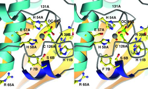
Figure 2.
A stereoview of the Zn-ligand cluster and the putative active site of LuxS. Helix α1 bearing the HXXEH motif is at the Left; the chain from A118 to A132 underpins the zinc-binding site, covering the Zn ion (white) and its ligands. Invariant residues are drawn in ball-and-stick mode; A and B designate the two chains of the dimer. The conserved Asp-37 of the B chain lies beneath Arg-39 and is not labeled; Asn-44 is above Arg-39B at the border of the picture. The N-terminal 310 helix that may control entry to the active site is dark blue. In this view the substrate-binding cavity lies below the water bound to zinc and nestles against the strands of the β-sheet from the B chain, seen at the back. Important interatomic distances in the active site region are as follows: Cys-84 Sγ—Zn, 4.86 Å; Arg-39 NH1—Cys-84 Sγ, 4.04 Å; Glu-57 Oɛ2—Zn, 4.69 Å.

