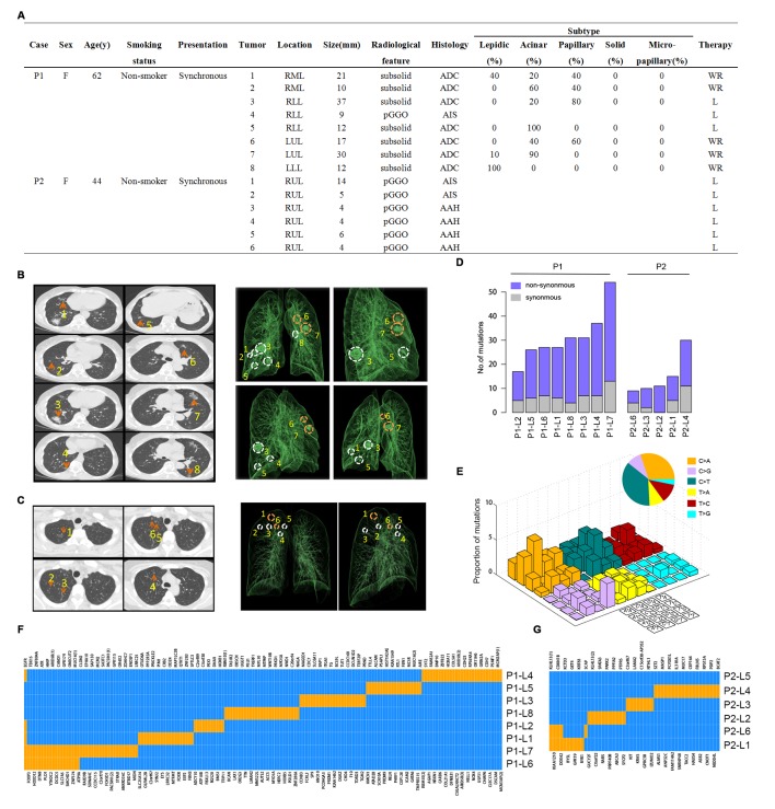
Figure 1.
Radiological features and whole-exome sequencing of the two patients. (A) Clinicopathological characteristics. RML, right middle lobe; RLL, right lower lobe; LUL, left upper lobe; LLL, left lower lobe; RUL, right upper lobe; pGGO, pure ground-glass opacities; ADC, adenocarcinoma; AIS, adenocarcinoma in situ; AAH, atypical adenomatous hyperplasia; WR, wedge resection; L, lobectomy; (B) Left: chest CT scans obtained with 1 mm thick sections of patient 1 (P1) with eight scattered GGOs (orange arrows) in the bilateral lung. Right: reconstructed lung 3D images of P1. Circles denote the spatial locations of the lesions and orange circles indicate metastatic lesions. (C) Left: CT scans obtained with 1 mm thick sections of patient 2 (P2) with six scattered GGOs (orange arrows) in her right upper lobe. All six lesions are pure GGOs of very small size. Right: reconstructed 3D images of P2. Circles denote the spatial locations of the lesions and orange circles indicate metastatic lesions. (D) Number of somatic mutations identified in each lesion of the two patients. Lesion name is in form of ‘Patient ID + lesion number’. For example, P1-L1 stands for the number 1 lesion of patient 1. (E) Mutational signature of P1 based on all somatic mutations detected in this patient. (F) Regional distribution of non-synonymous somatic mutations among the eight lesions of P1. Each column represents a single mutation site. Blue represents wild type while orange represents mutation in a certain site of a certain gene. (G) Regional distribution of somatic mutations among the six lesions of P2. Each column represents a single mutation site. Blue represents wild type while orange represents mutation in a certain site of a certain gene.

