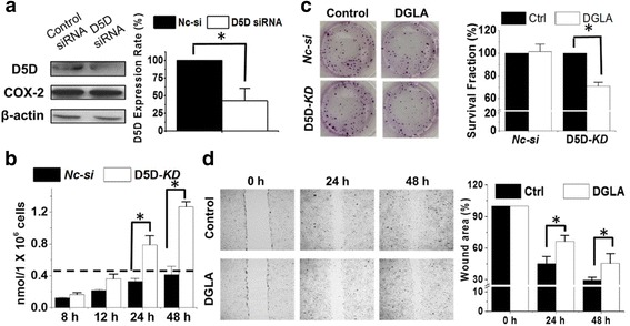
Fig. 3.

D5D-KD and DGLA inhibit 4 T1 cells growth and migration. a Western blot and protein expression rate of D5D and COX-2 in 4 T1 cells after D5D siRNA transfection (β-actin as loading control). Similar inhibition of D5D was also observed in shRNA transfected 4 T1 cells; b GC/MS quantification of 8-HOA from cell medium containing 1.0 × 10⁶ D5D-KD 4 T1 cells or control siRNA transfected cells after 100 μM DGLA treatment, Similar results were also observed in shRNA transfected cell lines vs. their controls (data not shown); c Colony formation of D5D-KD 4 T1 or control siRNA transfected cells at 10 days after DGLA treatment (100 μM for 48 h); and d Wound healing assays and quantification of wound area of D5D-KD 4 T1 cells upon DGLA (100 μM, 48 h) treatment vs. controls (without DGLA). Data represent as mean ± standard deviation (*: significant difference with p < 0.05 from n ≥ 3)
