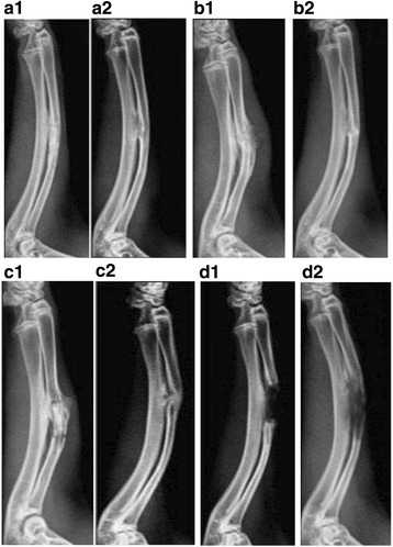
Fig. 4.

Radiographic examination of repair of bone defects. A1, A2 In ADSCs osteogenesis group, the bones were well connected, a marrow cavity appeared, and the bone defect was almost impossible to detect. B1, B2 In the ADSCs group, the bone marrow cavities became interconnected, but the defect was still obvious. C1, C2 In the negative control group, fracture lines were clear and the marrow cavity did not appear. D1, D2 In the blank control group, the bone defect was still visible. A1, B1, C1, D1: 4 weeks post-surgery; A2, B2. C2, D2: 8 weeks post-surgery
