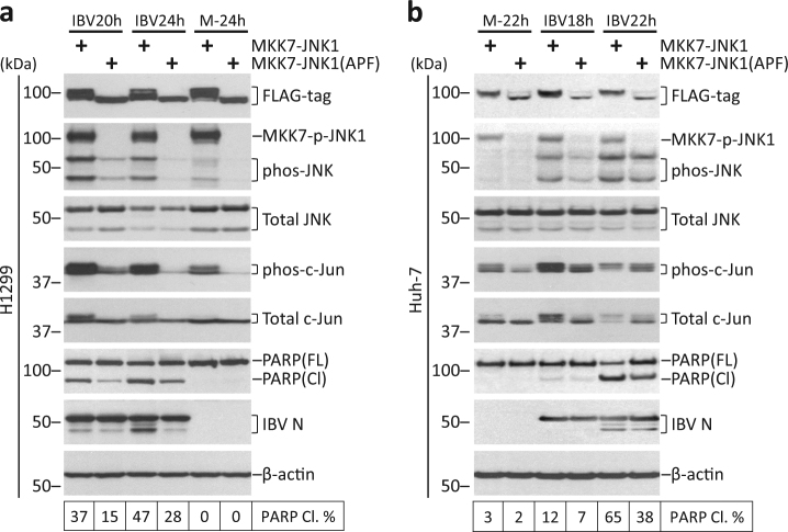
Fig. 4. Overexpression of constitutively active JNK promotes IBV-induced apoptosis.
a H1299 cells in duplicate were transfected with pcDNA-MKK7-JNK1 or pcDNA-MKK7-JNK1(APF), before being infected with IBV at MOI~2 or being mock infected. One set of cells were harvested for protein at the indicated time points and were subjected to Western blot analysis using the indicated antibodies. Beta-tubulin was included as loading control. Sizes of protein ladders in kDa were indicated on the left. Degree of JNK phosphorylation and the percentage of PARP cleavage was determined as in Fig. 3b. The experiment was repeated three times with similar results, and the result of one representative experiment is shown. b Huh-7 cells were transfected and infected similarly as in a. Western blot analysis and data quantification were performed as in a. The experiment was repeated three times with similar results, and the result of one representative experiment is shown.

