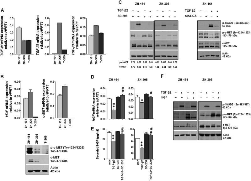
Fig. 1. Control of c-MET activity by TGF-β signaling.
a, b GIC were assessed for TGF-β 1/2/3 (a) and HGF, c-MET (b) mRNA levels by RT–PCR. Equal amounts of cellular lysates were assessed also for c-MET and p-c-MET levels by immunoblot (b). c GIC were seeded in complete NB medium in the presence of SD-208 (1 µM) or TGF-β2 (2 ng/ml) or both, or ALK-5 siRNA (100 nM) with or without TGFβ2 (2 ng/ml) for 24 h. DMSO diluted in NB medium (1:20’000) served as a control. p-SMAD2, p-c-MET and c-MET protein levels were assessed by immunoblot; 30 µg/lane protein for ZH-161 and 50 µg/lane for ZH-305 were loaded. Actin was used as loading control. Quantification of band intensity by ImageJ is shown below the immunoblot panels. d, e Modulation of HGF mRNA expression (d) and HGF protein release into the supernatant (e) by TGF-β2 stimulation in the absence or presence of SD-208 was assessed by RT–PCR and by ELISA (*p < 0.05, **p < 0.01, effect of TGF-β2 compared to control, # p < 0.05, ## p < 0.01 effect of TGF-β2 and SD-208 co-treatment compared to TGF-β2 alone). f The interference of TGF-β2 (2 ng/ml) with HGF (50 ng/ml) stimulation of c-MET phosphorylation was assessed by immunoblot

