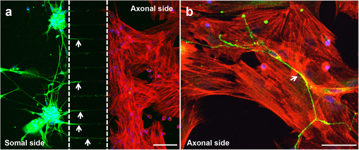
Fig. 2. Neurites cross the microgrooves to the axonal side of the microfluidic device.
Sensory neurons derived from rat DRG (5 × 10⁴ cells/cm2) and rat bone marrow MSCs (104 cells/cm2) were cocultured in microfluidic devices for 7 days. DRG neurons were maintained in DMEM supplemented with 2% (v/v) B-27 and 1 μM AraC; MSCs were incubated in OIM composed of DMEM-low glucose with 10% (v/v) FBS, 1 × 10⁻⁹ M dexamethasone, 10 mM β-glycerophosphate, and 50 μg/mL ascorbic acid. (a and b) The presence of neurites reaching MSCs was evaluated on day 7 of coculture by IF using an antibody directed against a neuronal specific marker (β-III Tubulin) coupled to Alexa Fluor® 488 (green), and DAPI (nuclei; blue) under a confocal microscope. Actin filaments of MSCs were stained using Alexa Fluor® 568 (red)-conjugated phalloidin. Arrows point to neurites. Scale bar = 100 µm a, 50 µm b

