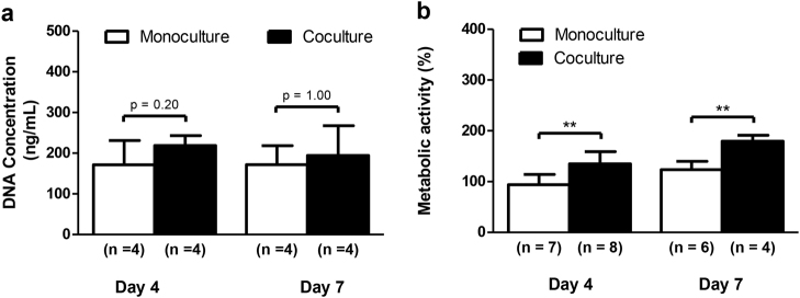
Fig. 3. DRG neurons stimulate the metabolic activity in MSCs.
Sensory neurons derived from rat DRG (5 × 10⁴ cells/cm2) and rat bone marrow MSCs (104 cells/cm2) were cocultured in microfluidic devices for 7 days. DRG neurons were maintained in DMEM supplemented with 2% (v/v) B-27 and 1 μM AraC; MSCs were incubated in OIM composed of DMEM-low glucose with 10% (v/v) FBS, 1 × 10⁻⁹ M dexamethasone, 10 mM β-glycerophosphate, and 50 μg/mL ascorbic acid. a The DNA concentration of MSCs was determined at 4 and 7 days of coculture by CyQUANT™ Cell Proliferation Assay. b The relative metabolic activity of MSCs was measured at 4 and 7 days of coculture by resazurin-based assay and normalized to the monoculture levels on day 4. Data expressed as mean ± SD. (n) indicates the total number of samples for each group. **p < 0.01 statistically different from monoculture. The results represent three independent experiments.

