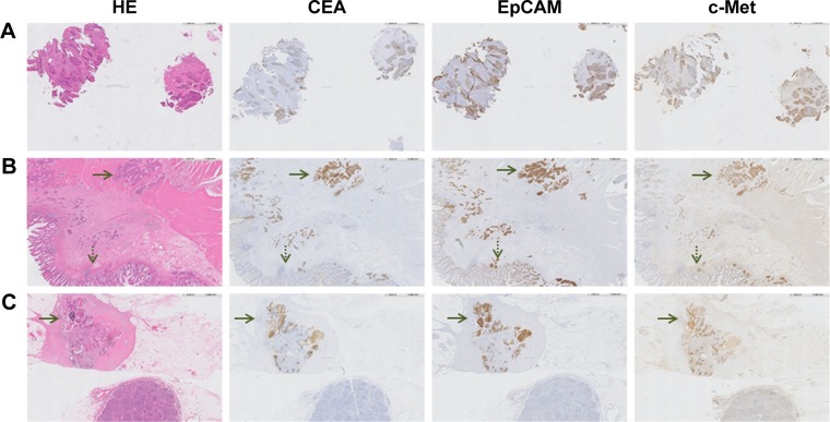
Figure 3.
Representative images of CEA, EpCAM and c-Met expression on RC tissues of a patient who was treated with neoadjuvant CRT. (A) Biopsy specimen (magnification ×5) obtained prior to the start of CRT, showing expression of all three biomarkers. (B) Primary tumor specimen (magnification ×5). The dotted arrow indicates normal epithelium and the other arrow indicates tumor tissue. A difference in intensity between tumor and normal tissue can be seen for all three biomarkers. This difference appears the highest for CEA. (C) Metastatic lymph node (magnification ×5). The arrow indicates the location of cancer cells, which are visualized by CEA, EpCAM and c-Met staining.
Abbreviations: HE, hematoxylin–eosin; CEA, carcinoembryonic antigen; EpCAM, epithelial cell adhesion molecule; c-Met, tyrosine-protein kinase Met; RC, rectal cancer; CRT, chemo- and radiotherapy.

