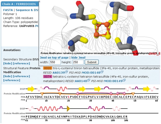
Fig. 2.

Protein modifications mapped onto the sequence and structure of ferredoxin I (PDB ID 1GAO, Chen et al., 2002). The Protein Modification track highlights residues involved in two iron-sulfur clusters (3Fe-4S (F3S): triangles/lines and 4Fe-4S (SF4): diamonds/lines). The number of edges of the protein modification icon symbolizes the number of residues involved in the modification. The 4Fe-4S cluster is displayed in the Jmol structure window above the sequence display
