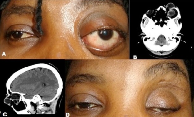
Figure 1.

Clinical and CT scan pictures of the patient
A: Pre-operative clinical picture of the patient showing left non-axial proptosis
B: CT scan (axial cut) picture of the patient showing the isodense cystic mass
C: CT scan (sagittal cut) of the patient showing the retrobulbar mass and a loculated mass within it
D: Post-operative clinical picture of the patient with severe ptosis
