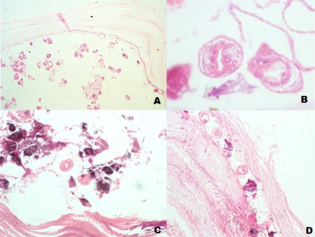
Figure 2.

Photomicrograph of the histology slides of the patient
A: Low power magnification of the germinal epithelium of the hydatid cysts giving off brood capsules and protoscoleses (×40)
B: Higher power magnification showing details of the protoscolex with hooks and suckers (×400)
C and D: Show dystrophic calcification of proscoleses and stromal fibrosis (×100)
