Figure 6.

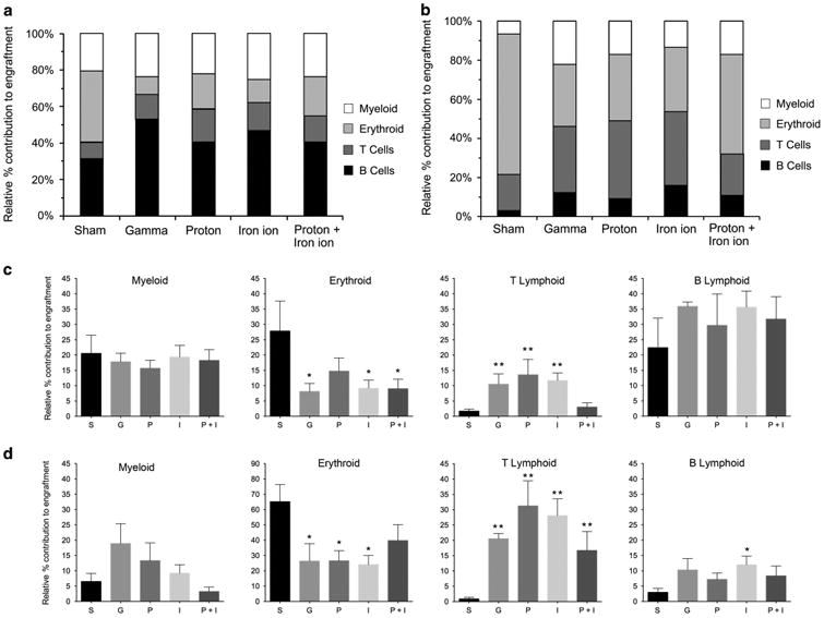
Effect of proton, iron ion and mixed-field proton/iron ion irradiation on differentiation/lineage commitment of human HSC in bone marrow or spleen of NSG mice: To assess the in vivo impact direct exposure of human HSC to mission-relevant doses of simulated SEP and/or GCR radiation has on differentiation/lineage commitment, human BM-derived CD34+ cells were exposed to the following: (1) monoenergetic protons (1 Gy, 50 MeV); (2) 56Fe ions (20 cGy, 1 GeV/n); or (3) sequential protons followed 15 min later by 56Fe ions at NSRL. Upon return to WFIRM, they were transplanted intravenously into busulfan-conditioned NSG mice. Mice were euthanized at 5–7 months post transplant, and then their long bones were collected and homogenized, and their spleens were collected and homogenized. Each were analyzed by flow cytometry with a panel of human-specific antibodies to CD antigens to identify various hematopoietic lineages within the recipient mice. (a) Relative lineage distribution of human cell engraftment in BM of recipient mice. (b) Relative lineage distribution of human cell engraftment in spleen of recipient mice. (c) Detailed analysis of lineage distribution of human cell engraftment in BM in response to each irradiation scheme. Data are displayed as mean ± s.e.m. (d) Detailed analysis of lineage distribution of human cell engraftment in spleen in response to each irradiation scheme. Data are displayed as mean ± s.e.m. For statistical analyses in c and d, lineage engraftment with human HSC exposed to each irradiation scheme was compared with lineage engraftment with sham-irradiated human HSC from the same donor. *P < 0.05; ** P < 0.01. Data outliers were removed for analysis in GraphPad Prism 6 setting Q = 2%.
