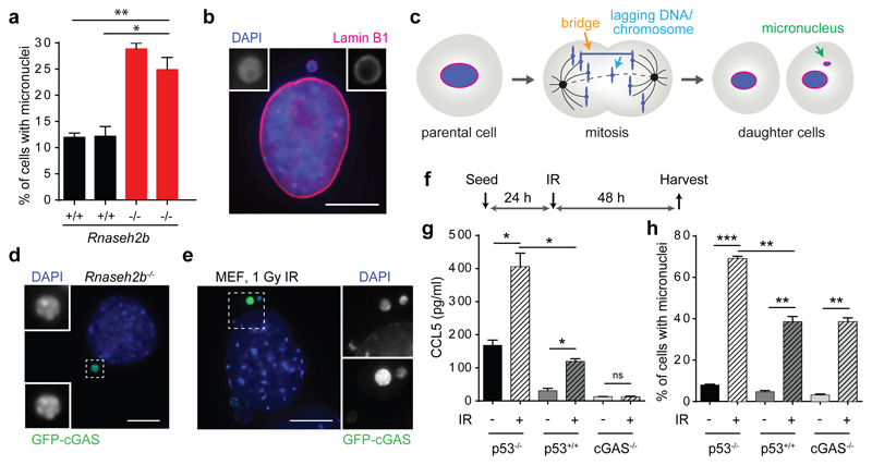
Fig 1. cGAS localises to micronuclei resulting from endogenous and exogenous DNA damage.
(a) Micronuclei form frequently in Rnaseh2b-/- MEFs, associated with genome instability. Percentage of cells with micronuclei in 2 Rnaseh2b+/+ control and 2 Rnaseh2b-/- MEF lines. Mean ± SEM of n=3 independent experiments (≥500 cells counted per line). (b) Micronuclear DNA is surrounded by its own nuclear envelope. Representative image with Lamin B1 (red) staining the nuclear envelope and DAPI staining DNA (blue). (c) Micronuclei form after mitosis as a consequence of impaired segregation of DNA during mitosis, originating from chromatin bridges and lagging chromosomes/chromatin fragments (further description, Supplementary Text). (d) GFP-cGAS localises to micronuclei in Rnaseh2b-/- MEFs. Representative image of GFP-cGAS expressing Rnaseh2b-/- MEFs. (e-h) cGAS localises to micronuclei induced by ionising radiation, and is associated with a cGAS-dependent proinflammatory response. (e) Representative image of GFP-cGAS positive micronuclei following 1 Gy IR in p53-/- MEFs. (f) p53-/-, p53+/+ and cGAS-/- MEFs were irradiated (1 Gy), and CCL5 production (g) and percentage of cells with micronuclei (h) assessed at 48 h. Mean ± SEM of n=2 independent experiments. * = P<0.05, ** = P<0.01, *** = P<0.001, two-tailed t-test; ns = not significant. Scale bars, 10 µm. Rnaseh2b+/+ and Rnaseh2b-/- MEFs in this figure and subsequent figures, are on a p53-/- C57BL/6J background (absence of p53 is a prerequisite for generation of Rnaseh2b-/- MEFs (20)).

