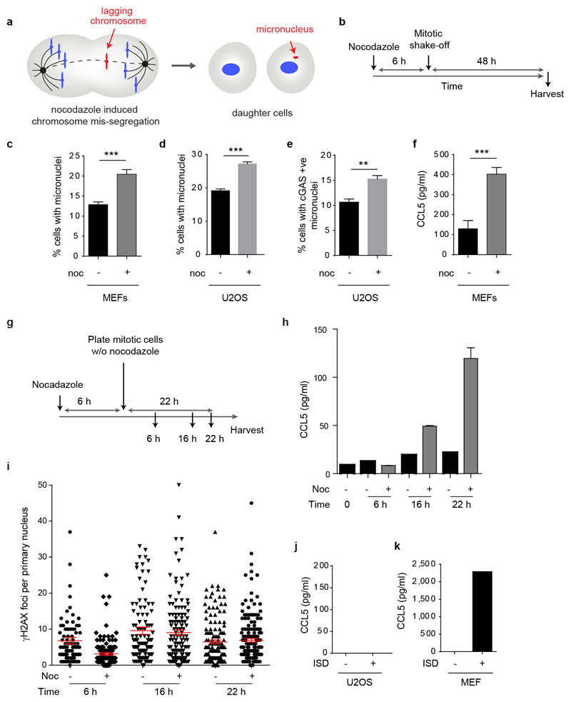
Extended data Fig 8. Induction of micronuclei originating from lagging chromosomes leads to a proinflammatory response, but not increased DNA damage in the primary nucleus.
(a) Model: micronuclei formation after nocodazole treatment. (b) Schematic of experimental protocol. (c) Percentage of micronucleated cells following nocodazole (noc) treatment of p53-/- MEFs or (d) U2OS cells. Mean ± SEM, n=5 experiments for p53-/- MEFs, n=3 for U2OS cells (e) Percentage of U2OS cells with cGAS positive micronuclei following nocodazole treatment. Mean ± SEM, n=3 experiments. c-e, ≥500 cells counted per experiment. (f) CCL5 secretion following nocodazole treatment of p53-/- MEFs. Mean ± SEM of n=5 experiments. ** = p<0.01, *** = p<0.001, two-tailed t-test; ns = not significant.(g-i) Increased CCL5 production after nocodazole release is observed after 16 h and not associated with increased DNA damage in the primary nucleus. (g) Experimental setup: p53-/- MEFs were arrested with nocodazole for 6 h, mitotic cells harvested by mitotic shake-off and re-plated in fresh media with nocodazole omitted. Supernatants and cells were then collected at indicated time points after growth in medium. (h) Increased CCL5 production was observed from 16 h after release from nocodazole block. Technical duplicate, mean ± SD. Noc (-), asynchronously grown, plated at the same time as mitotic shake-off Noc (+) cells, arrested with nocodazole. (i) No increase in numbers of γH2AX foci in the primary nucleus was observed after release from nocodazole block. n≥100 cells counted per condition. (j, k) CCL5 response to interferon stimulatory DNA (ISD) is absent in (j) U2OS cells but (k) present in MEFs. CCL5 measured by ELISA 8 h after transfection with ISD. n=2 experiments for U2OS cells, n=1 experiment for MEFs.

