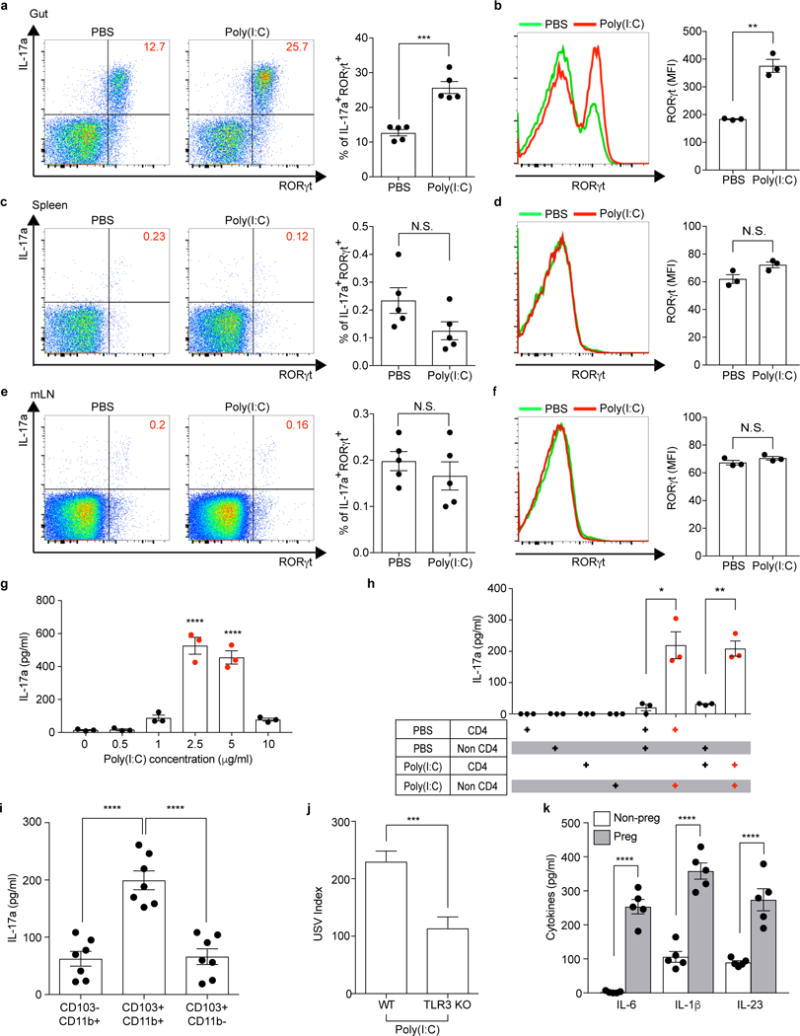
Extended Data Figure 6. CD11c+ cells stimulate gut-Th17 cells to produce high levels of IL-17a ex vivo.

a-f, Flow cytometry of CD4+ T cells (gated on TCR-β+CD4+) stained intracellularly for IL-17a and RORγt. Mononuclear cells were collected at E14.5 from the gut ilea, spleens, and mesenteric lymph nodes (mLN) of PBS-/poly(I:C)-treated mice (n=5/group (a, c, e); n=3/group (b, d, f)). MFI denotes mean fluorescence intensity. g-i,, Supernatant concentrations of IL-17a from mononuclear cells of the ilea in poly(I:C)-treated Tac dams (g) (n=3/group), from co-cultures of CD4+ and non-CD4+ cells of the ilea in PBS-/poly(I:C)-treated Tac dams (h) (n=3/group), or from co-cultures of CD4+ and CD103−CD11b+/CD103+CD11b+/CD103+CD11b− (gated on MHCII+CD11c+) cells of the ilea in poly(I:C)-treated dams (i) (n=7/group). All cultures were isolated at E14.5 and stimulated ex vivo with poly(I:C) for 18hrs (g-h) or for 48hrs (i). Data are pooled from 2 (g-h) or 3 (i) independent experiments. j. USV index (n=16/17 (poly(I:C);WT/TLR3 KO); 2 independent experiments). k, Supernatant concentrations of IL-6, IL-1β, and IL-23 from cultures of CD11c+ isolated at E14.5 from the ilea of poly(I:C)-treated non-pregnant/pregnant mice (n=5/group; 3 independent experiments). *p<0.05, **p<0.01, ***p<0.001 and ****p<0.0001 as calculated by Student’s t-test (a-f,j,k) and one-way ANOVA (g-i) with Tukey post-hoc tests. N.S., not significant. Graphs indicate mean +/− s.e.m.
