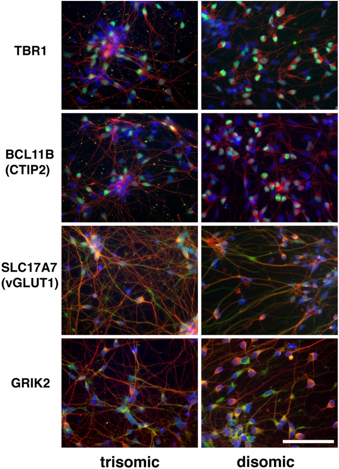
Fig 1. Confirmation of differentiation of iPSCs into cortical neuronal cultures.
Images taken from clones C2 (trisomic) and C3 -D21 (disomic) 40 days after initiation of the differentiation protocol. Fixed cells on chamber slides were probed with antibodies against the marker proteins listed in the left column. Neuronal marker Beta III tubulin is red; DAPI is blue; and Ctip2, TBR1, SLC17A7 (vGLUT1) and GRIK2 are green. Size bar = 20 μ.

