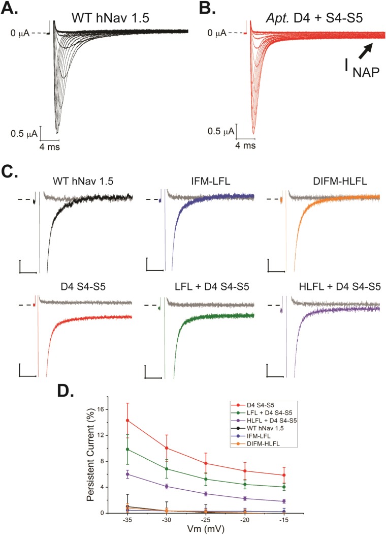
Fig 4. Apteronotini-specific Nav1.4b1 (scn4ab1) substitutions generate a persistent current in the human cardiac Nav1.5.
(A) WT hNav1.5 currents elicited by depolarizations in 5 mV steps from −80 mV to +15 mV. (B) Currents generated by an hNav1.5 variant with all 5 Apteronotini-specific substitutions (see Fig 3) in the D4 S4–S5 linker. Note the persistent (non-inactivating) current at the end of the pulse. (C) Normalized exemplar traces of late currents for hNav1.5 mutants. To facilitate comparisons among variants, current responses to steps to −60 mV (gray) and −20 mV (colors) are shown. Scale bars are 5% of peak current (vertical) and 5 milliseconds of time (horizontal), and dashed lines indicate the point of 0 current. (D) Quantification of percentage of persistent current for each variant over a range of membrane potentials. Error bars are ± 1 standard deviation. N ≥ 5 for each variant, in S4 Table. For all D4 S4–S5 substitution-containing variants, percentage of persistent current was significantly different (p < 0.01) as compared to WT hNav1.5. Figure data included in S2 Data. D4 S4–S5, R1644W/L1647W/M1651R/I1660F/G1661S; DIFM-HLFL, D1484H/I1485L/M1487L; hNav, human voltage-gated sodium; IFM-LFL, I1485L/M1487L; WT, wild-type.

