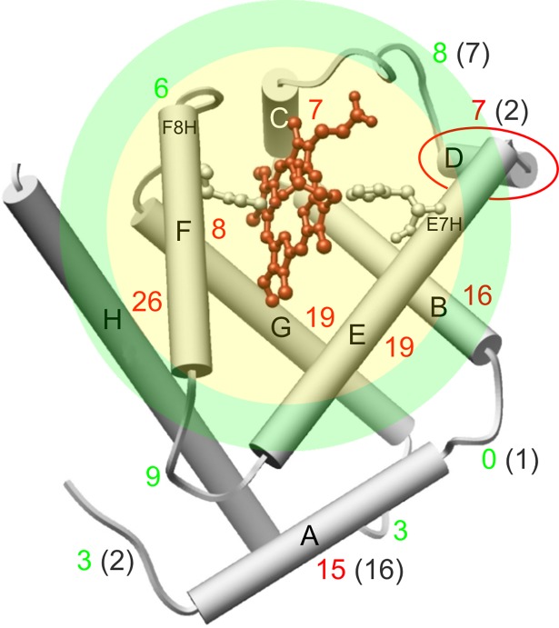
Fig 2. Structure of β subunits of hemoglobin (β-HbA).
α-helices are shown as cylinders, number of amino acids of segments is marked with colored digits: red for α-helices, green for loops (gray in the brackets refers to α subunit). The heme group is stabilized by the distal (E7H) and proximal (F8H) histidines. Helix D is marked with red ellipse as the dominant difference between β and α subunits [6]. The transparent colored discs denote the estimated vicinity of the heme of which is characterized by the compressibility in the present work. Yellow and green area mean the spheres with radius R98% and R99%, labelling a rage of estimation with 2% and with 1% error respectively (see Appendix II).

