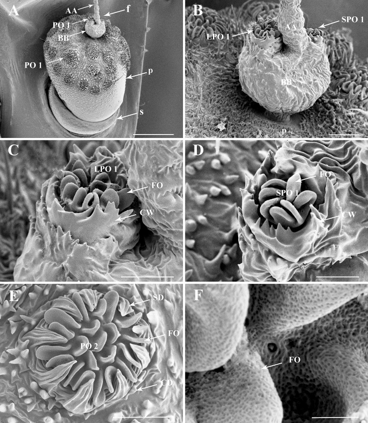
Fig 4. Details of the antenna of the third nymphal instar of Lycorma delicatula.
(A) General view of the antennae, showing the distribution of sensilla on antennal surface. (B) Basal bulb of antennal flagellum, LPO 1 and SPO1. (C) LPO 1 on the anterior faces of the antenna. (D) SPO1 on the posterior face of the antenna. (E) PO 2 of the pedicel. (F) Multiporous surface of PO2 lobes. Scale bars: A = 150 μm; B = 25 μm; C = 15 μm; D = 10 μm; E = 20 μm; F = 2 μm. Abbreviations: AA, apical arista of antennal flagellum; BB, basal bulb of antennal flagellum; CD, cuticular denticles; CW, encircling walls; f, flagellum; FO, multiporous folds; p, pedicel; PO1, plaque organ type I; PO2, plaque organ type II; s, scape; SD, sclerotized denticles.

