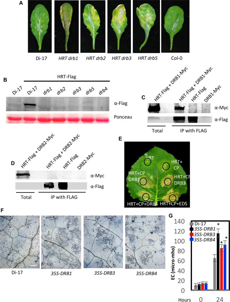
Fig 4. DRB proteins are required for the stability of HRT.
(A) HR formation in TCV-inoculated Di-17, Col-0 and HRT drb genotypes at 10 dpi. The HR phenotype was evaluated in ~20–30 plants that were analyzed in four separate experiments. (B) Western blots showing relative levels of HRT-Flag in Di-17 and drb genotypes expressing HRT-Flag transgene. Ponceau-S staining of the Western blots was used as the loading control. This experiment was repeated three times with similar results. (C and D) IP of DRB1-Myc (C) and DRB2-Myc (D) with HRT-Flag. All proteins were expressed under their respective native promoters in Arabidopsis. The immunoprecipitated proteins were analyzed with α-Myc and α-Flag and this experiment was repeated twice with similar results. (E) Visual phenotype of Nicotiana benthamiana leaves expressing indicated proteins. Agroinfiltration was used to express HRT, CP, EDS1 (E90-At3g48090), and DRB1, DRB3 or DRB4 proteins. The leaf was photographed at 4 days post treatment. (F) Trypan blue stained leaves of Di-17 and transgenic plants overexpressing DRB1, DRB3 and DRB4 in Di-17 background. The plants were inoculated with TCV and the inoculated leaves were sampled at 36 h post inoculation. Scale bars, 270 microns. At least four independent leaves were analyzed with similar results. (G) Electrolyte leakage in genotypes shown in F. The leaves were sampled at 0 and 24 h post TCV inoculation. Error bars represent SD. Asterisks indicate data statistically significant from that of control (Col-0) (P<0.05, n = 4).

