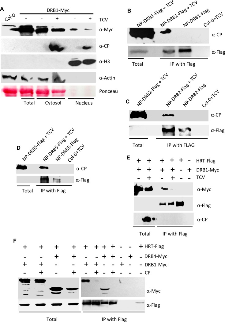
Fig 5. DRB proteins interact with CP.
(A) Relative levels of DRB1 in nucleus and cytosolic fractions of Arabidopsis plants expressing DRB1-Myc under its self promoter. The blot was sequentially probed with indicated antibodies. Ponceau-S staining of the Western blot was used as the loading control. This experiment was repeated two times with similar results. Fold change, normalized with Rubisco, Actin or H3 proteins, in western blots was quantified using Image Quant software. (B-D) Co-IP of DRB1-Flag, (B) DRB2-Flag (C) and DRB5-Flag (D) in the presence of TCV. The transgenic Arabidopsis plants expressing DRB1-Flag, DRB2-Flag, and DRB5-Flag under their respective native promoters (NP) were inoculated with TCV and leaves sampled at 3 dpi were processed for Co-IP. The TCV inoculated Col-0 plants were used as a negative control. These experiments were repeated twice with similar results. (E and F) Co-IP of DRB1-Myc or DRB4-Myc with HRT-Flag in the presence or absence of TCV (E) or CP (F). Arabidopsis expressing DRB1 and HRT under their native promoters were used in E. For transient assays shown in F, N. benthamiana plants were agroinfiltrated and immunoprecipitated proteins were analyzed with α-Myc and α-Flag. The experiments shown in E and F were repeated two times with similar results.

